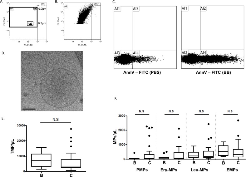
Figure 1. Microparticles in pleural effusions.
A. Flow cytometry scattergram of the microparticle (MP) window of analysis determined by FSC-Megamix-Plus beads. B. Representative scattergram of the pleural fluid events appearing in the MP window. C. Representative dot plot showing the annexin-V (AnnV) positivity of the pleural fluid extracellular vesicles. The control experiment was performed in the presence of phosphate buffered saline buffer (PBS) compared to Ca2+-containing binding buffer (BB). D. Representative image of pleural fluid extracellular vesicles by cryo-transmission electron microscopy. E. Total MP counts by flow cytometry (TMP = AnnV+MPs) between benign B. and cancer C. pleural fluids. F. Hematopoietic and vascular MP subpopulation enumeration by flow cytometry between benign B. and cancer C. pleural effusions. Platelet-derived MPs (PMPs): AnnV+/CD41+; erythrocyte-derived MPs (Ery-MPs): AnnV+/CD235a+; Leucocyte-derived MPs (Leu-MPs): AnnV+/CD11b+; endothelial-derived MPs (EMPs): AnnV+/CD41−/CD31+. NS = no significant difference.

