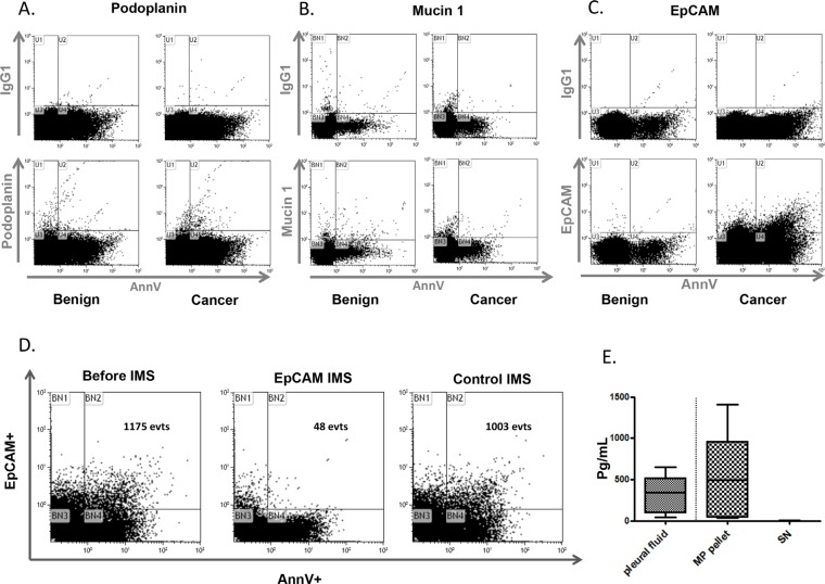
Figure 2. Tumoral microparticles in pleural effusion.
Representative flow cytometry graphs of podoplanin A. mucin 1 B. and EpCAM C. labeling on MPs from benign B. or cancer C. pleural fluids. The control experiments with appropriate isotype antibodies are displayed above each specific graph. D. The specificity of EpCAM+ microparticles in malignant pleural effusions. Representative experiment of AnnV+/EpCAM+MP labeling by flow cytometry before and after immunomagnetic separation (IMS) using beads coated with the EpCAM antibody. The control IMS was performed with beads coated with an irrelevant antibody. E. The EpCAM antigens are vectorized by MPs. Comparison of the EpCAM antigen determined by ELISA between the pleural fluids, MP pellets and last-wash supernatants (SN) (n = 5).

