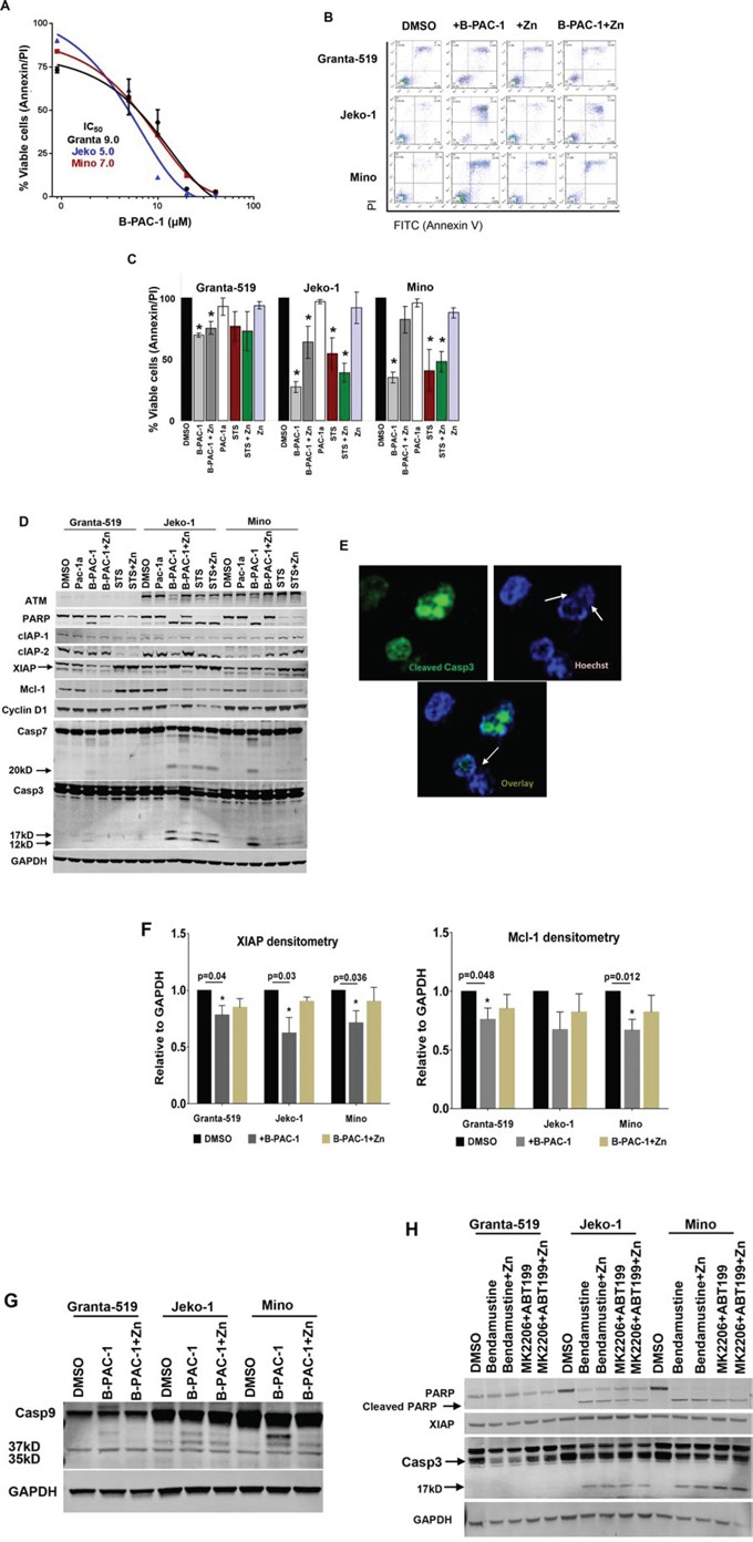
Figure 1. B-PAC-1 induce cell death in MCL cell lines by activating Casp3.

A. Dose dependent cell death by B-PAC-1 in MCL cell lines. Granta-519, Jeko-1 and Mino cells were incubated with increasing concentration of B-PAC-1 for 24 hr, stained with Annexin V-PI and acquired in FACS caliber for cell death analysis. Their respective IC50 value was determined as shown in the inset. B. FACS analysis showing differential cell death induced by B-PAC-1 (10 μM, 24 hr) with or without Zn in Granta-519, Jeko-1 or Mino cells. C. Annexin V-PI FACS analysis of B-PAC-1 induced cell death (Granta-519; *p < 0.0001; Jeko-1; *p < 0.0001 and Mino; *p < 0.0001) or inhibition by Zn (Granta-519; *p < 0.007; Jeko-1; *p < 0.035) (n = 5; Mean ± SE) as described in B. Pac-1a, was used as negative control while staurosporine (STS;100 nM) was used as positive control (n = 3 *p < 0.03–0.004 in Granta-519; *p < 0.03–0.002 in Jeko-1 or *p < 0.020 - 0.003 in Mino cells compared with DMSO control. D. Western blot analysis of protein extracts (30 μg) from Granta-519, Jeko-1 and Mino cells treated with indicated compounds for 24 hr showing cleavage of Casp3 and Casp7 by B-PAC-1 and STS accompanied by cleavage of both Casp3 substrates ATM and PARP and corresponding loss of XIAP, Mcl-1, cIAP-1 and cIAP-2 proteins. Treatment with inactive Pac-1a (10 μM) was used as negative control and Zn was utilized to abrogate B-PAC-1 induced PCD. GAPDH was used for loading control. Identical blots were either reprobed or cut in strips and separately probed with antibodies for indicated proteins. E. Immunofluorescence analysis of Jeko-1 cells treated with B-PAC-1 for 24 hr showing Casp3 cleavage is accompanied by nucleosomal pyknosis. Arrows indicate nuclear pyknosis in cleaved Casp3 expressing cells. F. Densitometry analysis (n = 4; Mean ± SE) showing loss of anti-apoptotic proteins XIAP and Mcl-1 following treatment with B-PAC-1 and Zn in Granta-519, Jeko-1 and Mino cells. *Significant difference from control. G. Western blot analysis (30 μg) of protein extracts from Granta-519, Jeko-1 and Mino cells showing cleavage of Casp9. Arrows indicating 37 and 35kD cleaved bands. GAPDH was used for loading control. H. Western blot (30 μg) analysis showing cleavage of Casp3 and PARP and loss of XIAP in MCL cell lines treated with Bendamustine (30 μM) or a combination of ABT199 (20 μM) and MK2206 (5 μM) for 24 hr in presence or absence of Zn (100 nM). GAPDH was used for loading control.
