Figure-10.

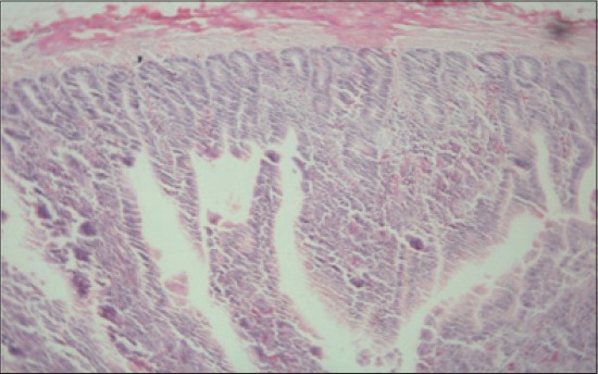
Jejunum from control group (T1) showing diffuse infiltration of inflammatory cells, predominantly heterophils and clubbing of villi (H and E, ×2.5).

Jejunum from control group (T1) showing diffuse infiltration of inflammatory cells, predominantly heterophils and clubbing of villi (H and E, ×2.5).