Fig. 4.
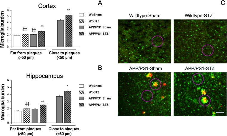
Microglial activation in STZ-treated mice. a Microglial burden was significantly increased in SP-free areas in the cortex from APP/PS1-Sham and Wt-STZ-treated mice. Moreover, this effect was worsened in APP/PS1-STZ-treated mice (**p = 0.01 vs. the rest of the groups, ‡‡p < 0.01 vs. Wt-Sham). Microglial burden in close proximity to SP was significantly higher in APP/PS1-STZ-treated mice (**p < 0.01 vs. APP/PS1-Sham). b A similar profile was observed in the hippocampus, where increased microglial burden in SP-free areas from Wt-STZ-treated mice was worsened in APP/PS1-STZ-treated animals (**p < 0.01 vs. the rest of the groups, ‡‡p < 0.01 vs. Wt-Sham). In the proximity of SP, microglial burden was significantly higher in APP/PS1-STZ mice (*p < 0.04 vs. APP/PS1-Sham). c Illustrative examples of cortical microglial immunostaining using anti-microglia (iba 1, green) and anti-Aβ (4G8, red) where increased microglial burden can be detected in APP/PS1-STZ mice, both in proximity to and far from SP. Scale bar = 125 μm

