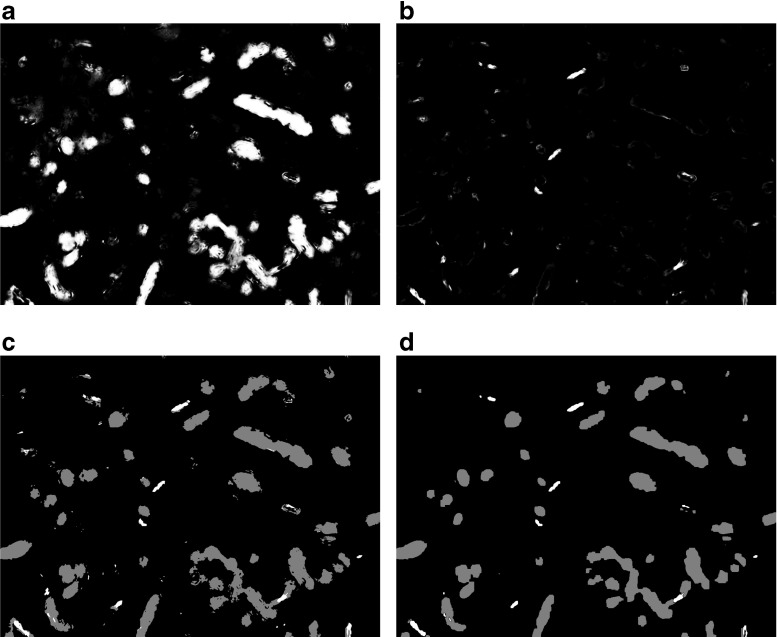
Fig. 3.
Classification and regularization of a single image. (a) and (b): Probability maps obtained by a Gaussian classifier that has been trained with the features extracted from the same image that is shown in Figure 2. The pixel-wise probability of belonging to the label ”Mitochondrion” is shown in (a) and the pixel-wise probability of belonging to the label ”Synaptic junction” is shown in (b). (c): preliminary segmentation before regularization, where each pixel has simply been given the label with highest probability (Mitochondria, gray; Synaptic junctions, white). Note the sparse pixels scattered throughout the image, the small holes in some of the segmented objects and the jagged edges. (d): Final segmentation after regularization via CRF energy minimization. Most sparse pixels have disappeared and edges show a smoother appearance

