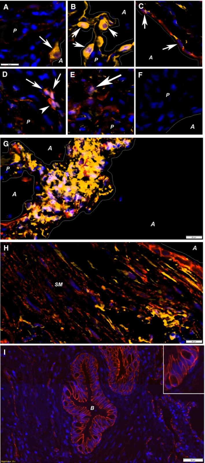
Figure 2.

Double stainings with β‐catenin. Several double stainings with β‐catenin was performed to investigate the cell types activated. These double stainings showed colocalization (arrows) of β‐catenin (red staining in all images) with Prosurfactant Protein C (A), pan‐cytokeratin (B), vimentin (C), NLK (D), and ICAT (E). These antibodies were all visualized with Cy3 (yellow labeling), and DAPI (blue) was used to visualize nuclei. Negative controls, omitting the secondary antibody (F) showed no positive labeling. β‐catenin/vimentin‐positive cells (purple nucleus in yellow cells) were also found in foci within border zones (G) as well as within smooth muscle bundles (H) in association with bronchial epithelium. Vimentin‐positive epithelial cells (C) suggest presence of EMT. Interestingly, β‐catenin was found both in cellular junctions/in the cell membrane (D, arrowhead and I) and within nuclei (arrows in A, B, C, D and E). All images, except D, E, and I are taken in border zones, whereas D and E are taken in densely fibrotic tissue where no alveolar structures were found and I shows a bronchiole. Structural elements within the images are marked; lines illustrate the border between parenchymal (P) tissue and alveolar (A) spaces, in addition to smooth muscle bundles (SM) and bronchioles (B). Scale bars represent 20 μm in A–H, 50 μm in I, and scale bar in A is applicable to images A–F.
