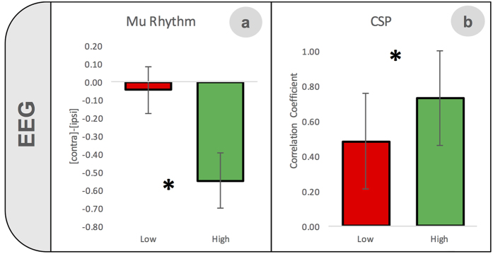
Figure 4. Significant differences between high- and low-aptitude BMI users in EEG-correlates of pure motor imagery.
The difference in μ-rhythm suppression (mean values ± standard error) between two clusters of electrodes, one over each hemisphere (index of laterality) was significantly more marked in the high- than in the low-aptitude group, showing the importance of a specific lateralized activation during MI for BMI control (a). The correlation (mean values ± standard error) between the right and left common spatial patterns (CSP) was higher in high- rather than low-aptitude users, indicating a more lateralized cortical activity during MI in the former group (b).

