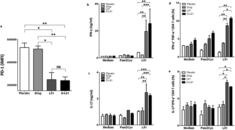
Figure 2. L91 immunization rescues CD4 T cells from exhaustion and activates polyfunctional Th1 cells in the drug treated and Mtb exposed mice.
At 20 wk of Mtb infection, lung cells from D-L91 treated mice were isolated and expression of PD-1 was examined by flowcytometery. (a) Bar diagram display integrated mean fluorescence intensity (iMFI) of PD-1 marker on CD4+ T cells. Lung cells were isolated from infected mice undergone combinatorial therapy of drug and L91 and single cell suspensions were prepared and in vitro stimulated with L91. Control cultures were incubated with Pam2Cys and medium. After 72 h, SNs of stimulated culture were examined for the secretion of (b) IFN-γ and (c) IL-17A by ELISA. The co-expression of IFN-γ/TNF-α and IL-17A/IFN-γ on CD4+ T cells was examined by flowcytometery. Bar diagrams illustrating CD4 T cells of lungs co-expressing (d) IFN-γ+TNF-α+ and (e) IL-17A+IFN-γ+. Data are representative of 2–3 experiments (n = 3 mice/group) and depicted as means ± SEM. D-L91: mice treated with combinatorial therapy of INH + RFP in drinking water along with L91. *P ≤ 0.05, **P ≤ 0.005, ***P ≤ 0.0005, ns = none significant. D-L91: mice administered with drugs in association with L91.

