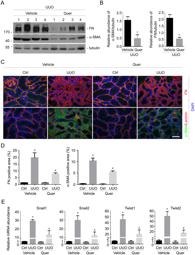
Figure 2. Quercetin reduces FN and α-SMA expression in the UUO kidneys.
(A) Western blot analyses showing the abundance of FN and α-SMA in the kidneys at 2 weeks after UUO w/o quercetin treatment. The gels were run under the same experimental conditions. (B) Graphic presentation showing FN and α-SMA protein abundance from (A) in the UUO kidneys. *P < 0.05 compared with UUO kidneys (n = 4). (C) Representative micrographs showing immuno-staining for FN and α-SMA expression in various groups as indicated. Scale bar = 50 μm. (D) Graphic presentation showing the quantitative results for FN and α-SMA immuno-staining in kidney tissue within groups. *P < 0.05 compared with contra lateral kidneys (n = 4), #P < 0.05 compared with UUO kidneys (n = 4). (E) Real time PCR analysis for Snail1, Snail2, Twist1 and Twist2 mRNA expression in the kidney tissue among groups. *P < 0.05 compared with contra lateral kidneys (n = 5), #P < 0.05 compared with UUO kidneys (n = 5).

