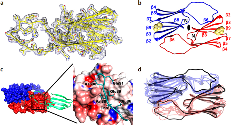
Figure 5.
(a) Electron density of AgsA:His-DD monomer, contoured at 1.0 σ. (b) Structure of the symmetric ACD dimer. Protomers are in red and blue, Methyl Pentanediol molecule in yellow. (c) Binding of dimerization loop (green) in β4–β8 groove of neighbouring molecule in lattice (red and blue surface) (Left). Close-up of boxed region, loop shown in green sticks, groove as electrostatic surface (Right). (d) Superposition of AgsA:His-DD dimer (black) with all other non-metazoan sHSP dimers.

