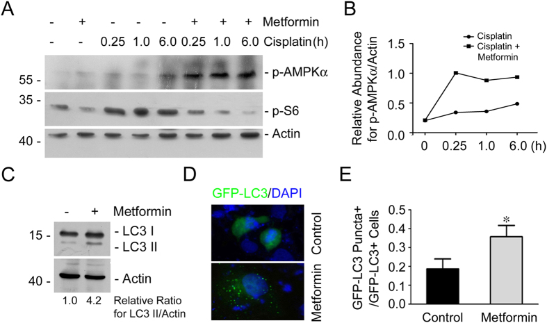
Figure 6. Metformin promotes AMPKα activation and autophagy induction in NRK-52E cells.
(A,B) Western blot analysis showing the abundance of p-AMPKα and p-S6 at different time points after cisplatin treatment in NRK-52E cells. The antibody against actin was probed as normalization. The gels were run under the same experimental conditions. (C) Western blot analysis showing the induction of LC3-II abundance in NRK-52E cells treated with metformin plus cisplatin. The gels were run under the same experimental conditions. (D) Representative micrographs showing the autophagosome formation in NRK-52E cells after metformin treatment. NRK-52E cells were transiently transfected with GFP-LC3 expression plasmid for 24 h, followed by metformin treatment for 12 h. The transfected cells with dot-like GFP-LC3 puncta were considered as autophagosome formation in the cells. (E) The graph showing the quantitative analysis for autophagosome formation in NRK-52E cells after metformin treatment. Data are presented as the percentage of the GFP-LC3 puncta positive cells. *P < 0.05 compared to the control cells (n = 3).

