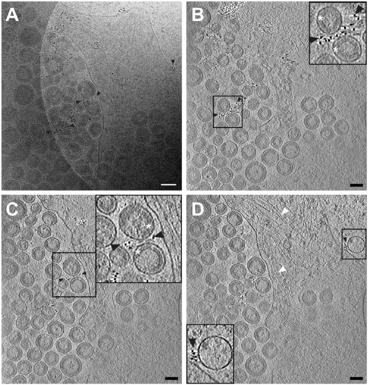
Figure 1.
Cryo-electron tomography of immunolabeled-tetherin and HIV-1 virus-like particles (VLPs) attached to an HT1080 cell. (A) The central projection image from a tilt series of HIV-1 VLPs tethered to the edge of an HT1080 cell. Image was low-pass filtered using ImageJ (Gaussian blur, Schneider et al., 2012). (B–D) Slices (7.64 nm) near the bottom (B), through the middle (C), and at the top (D) of the 3D reconstruction, showing a visible, ordered Gag lattice in the HIV-1 VLPs and immunogold-labeled tetherin located on the plasma membrane (D) and on HIV-1 VLPs (B–D), as indicated by black arrowheads. Cytoskeletal elements are visible, as indicated by white arrowheads in a 3D tomographic slice (D). Insets in (B–D) (black boxes) are 2× magnification. White asterisks indicate the immature HIV-1 Gag lattice in insets for panels (B) and (C). Gold fiducial markers, 20 nm in diameter, were added to the sample and used as image alignment aids during the 3D tomographic reconstruction process. Scale, 50 nm.

