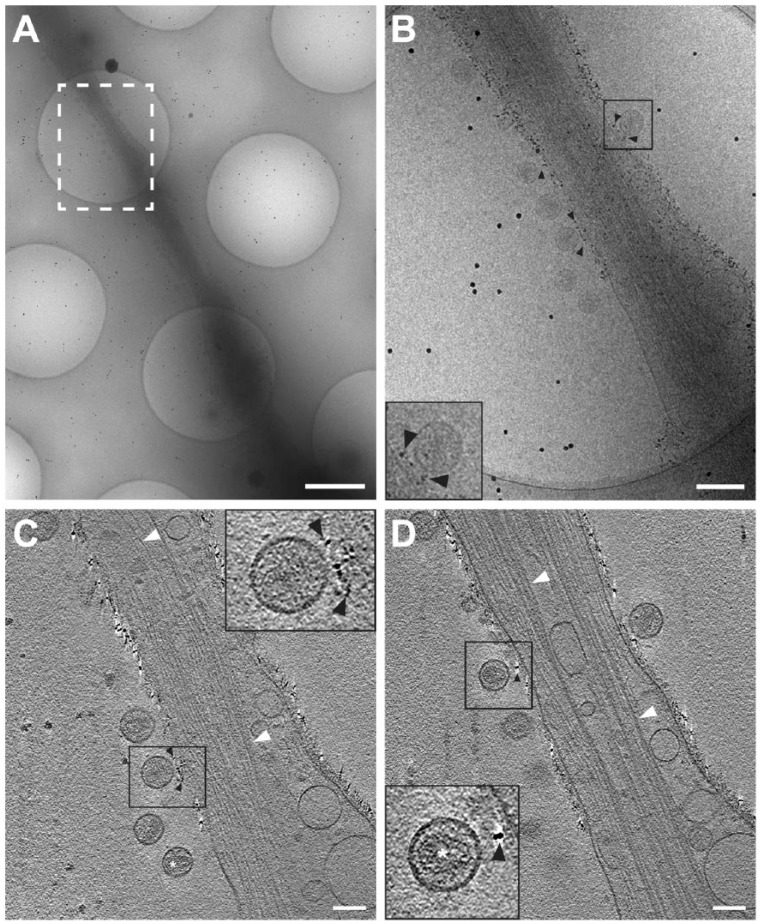
Figure 2.
Cryo-electron tomography (Cryo-ET) of immunolabeled tetherin and HIV-1 virions attached to an HT1080 cell microspike. (A) Cryo-TEM 2D image montage of HIV-1 tethered to a thin HT1080 cell extension. The dashed white box highlights the area of enlargement in (B). (C and D) Slices (8.84 nm) through the 3D reconstruction; black arrowheads indicate immunogold-labeled tetherin located between HIV-1 virions and the plasma membrane. Preserved cytoskeletal elements are visible in the 2D images and the 3D tomographic slices, as indicated by white arrowheads. White asterisk was placed over typical mature HIV-1 conical cores in panel (C) and the inset for panel (D). Insets in (B–D) (black boxes) are 2× magnification. Gold fiducial markers, 20-nm in diameter, were added to the sample and used as image alignment aids during the 3D tomographic reconstruction process. Scale (A) 500 nm; (A) and (B–D) 100 nm.

