Figure 5.
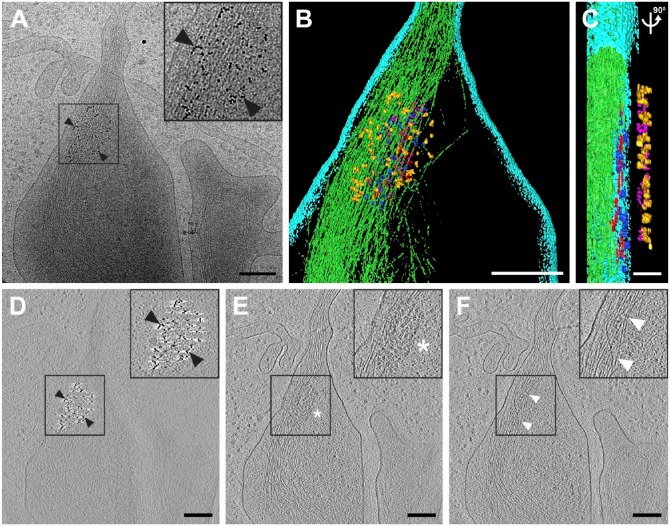
Early hRSV assembly site detected by native immunolabeling. (A) 2D cryo-electron microscopy image of an hRSV assembly site; the 6-nm gold particles indicate the location of the hRSV F glycoprotein (black arrowheads) at the upper surface of the plasma membrane. (B and C) Segmented 3D volume of the hRSV and cellular macromolecules. (B) Top view. (C) Cut-away and side view of (B) with 90° rotation applied. The cell membrane is presented in cyan. The filamentous actin network is noted in green. The matrix protein is depicted in blue. The glycoprotein densities are highlighted in magenta. Red tubular densities correspond to ribonucleoprotein (RNP). Gold densities are the 6-nm gold particles conjugated to the secondary antibody. (D–F) Slices (7.64 nm) through the reconstructed 3D volume showing the gold on the top (D, black arrowheads), a quarter-plane slice noting the hRSV viral proteins (E, white asterisk), and a bottom slice highlighting the actin filaments (F, white arrowheads). Inset in A is 2× magnification, insets in D-F are 1.5× magnification. Gold fiducial markers, 20 nm in diameter, were added to the sample and used as image alignment aids during the 3D tomographic reconstruction process. Scale (A, B, D–F) 200 nm; (C) 50 nm.

