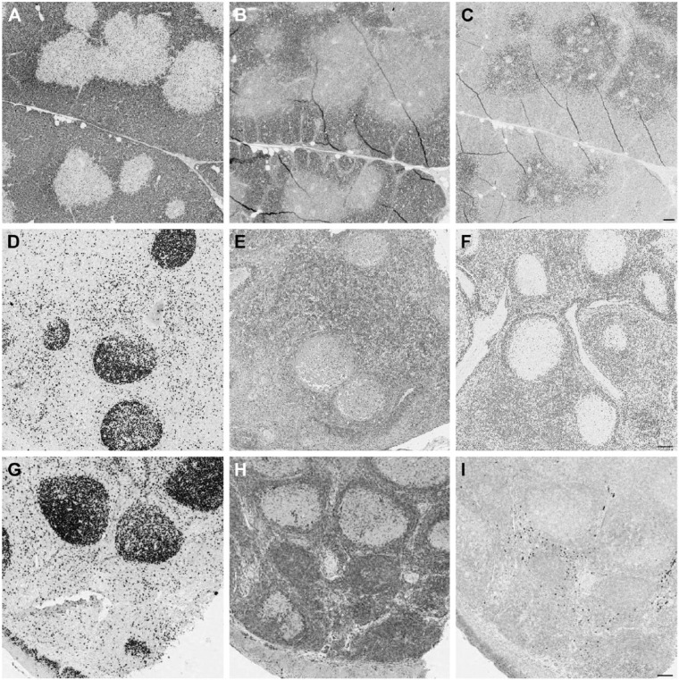
Figure 4.
CDKN1B (p27) antibodies stain opposite compartments in swine thymus. Porcine thymus (A, B and C), lymph node (D, E and F) and human tonsil (G, H and I) are stained with Ki-67 MIB 1 antibody (A, D and G), mouse anti-p27 (B, E and H) and rabbit anti-p27 (C, F and I). Note the opposite staining pattern of the mouse (B) and rabbit (C) reagents in the thymus. In the peripheral swine and human tissues, proliferating germinal center cells are unstained. Note the reversed staining intensity of both anti-p27 antibodies with porcine and human tissue. Double staining for Ki-67 and both anti-p27 antibodies showed that proliferating corticothymocytes were mutually exclusively labeled by the rabbit antibody and co-expressed CDKN1B with the mouse reagent; this co-expression is the expected distribution (Nagahama et al. 2001). Scale, 100 µm

