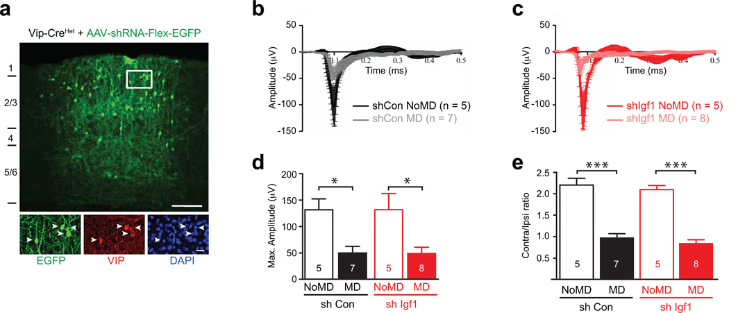
Extended Data Figure 8. VIP neuron-derived IGF-1 does not disrupt ocular dominance plasticity.
a) Widespread infection of VIP neurons by AAV-shRNA-hUbc-Flex-EGFP. High-titer injection of AAVs into the visual cortex of P18-20 Vip-Cre/+ mice leads to infection of the majority VIP neurons (green = EGFP, red = anti-VIP, blue = DAPI, arrowheads = infected VIP neurons; Scale bars 150 µm, 20 µm in the inlet).
b–c) Average of VEP traces recorded in the visual cortices of mice that were injected with AAVs expressing control shRNA (black/grey) or shRNA against Igf1 (red/pink) shRNA and that were (grey, pink) or were not (black, red) subjected to monocular deprivation in the eye contralateral to the recording site (MD vs NoMD, respectively).
d) Monocular deprivation induces a significant reduction in the VEP amplitude in response to low spatial frequency stimulation in mice that had AAVs expressing control shRNA and Igf1 shRNA injected into their visual cortices (control shRNA NoMD, n = 5 mice; control shRNA MD, n = 7 mice; Igf1 shRNA NoMD, n = 5; Igf1 shRNA MD, n = 8. p* < 0.05, Mann Whitney U-Test).
e) Mice that had AAVs expressing control shRNA (black) and Igf1 shRNA (red) injected into their visual cortices display normal ocular dominance plasticity as monocular deprivation (MD) induces a shift to the ispilateral eye in both groups (control shRNA NoMD, n = 5 mice; control shRNA MD, n = 7; Igf1 shRNA NoMD, n = 5 ; Igf1 shRNA MD, n = 8 mice. p** < 0.0001, One-way ANOVA-Tukey post test).

