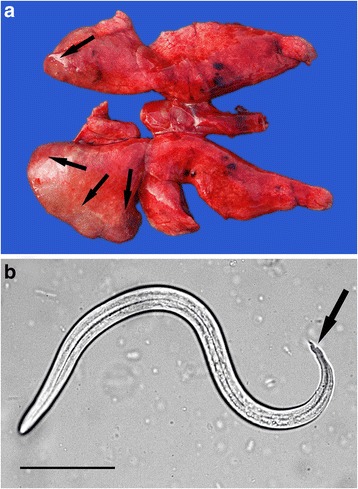
Fig. 5.

Angiostrongylosis in a stoat. a The lungs show areas of consolidation and swelling (arrows) along the ventral margins of the diaphragmatic lobes caused by Angiostrongylus vasorum infection. The areas of haemorrhage in the cranial lobes were a result of trauma caused by road traffic. b One of many first stage larva of A. vasorum seen in a wet impression taken from the cut surface of the lung. Note the wavy, double notched tail-tip which is characteristic of A. vasorum (arrow). Bar = 50 μm
