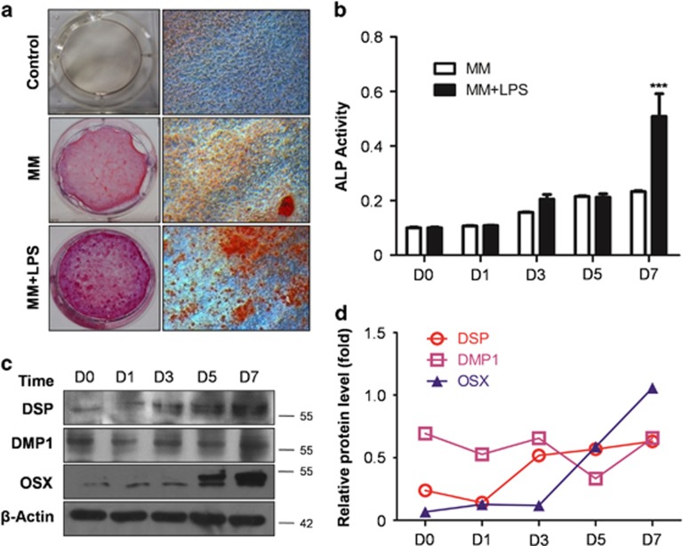
Figure 1.
LPS enhances odontoblast differentiation capability. (a) mDPC6T were cultured in completed medium (Ctrl), MM and MM added LPS (MM+LPS) for 14 days. Alizarin red S was used to stain mineralization with red in the dishes. There are few red mineralized nodules in control group, whereas scattered mineralized nodules can be seen in MM. More minerals were detected in MM+LPS compared with MM group. (b) ALP activity in MM and MM+LPS group at different time points. It showed no significant difference between two groups within 5 days, nevertheless ALP activity was upregulated obviously in MM+LPS group at 7 days. Mean±S.E.M.; ***P<0.001. (c) Odontoblasts differentiation markers were detected with DSP, DMP1 and OSX during the process of differentiation in MM+LPS. The protein level of DSP, DMP1 and OSX were increased time dependently within 7 days. (d) Protein levels of DSP, DMP1 and OSX in the MM+LPS group were quantitated using densitometry. The experiments were repeated in triplicate, we analyzed every result of the experiments and made sure they showed the same pattern. Each target protein was normalized to the β-actin levels to provide a visible tendency, so the curve showed a relative protein level. Experiments were repeated at least twice

