Figure 6.
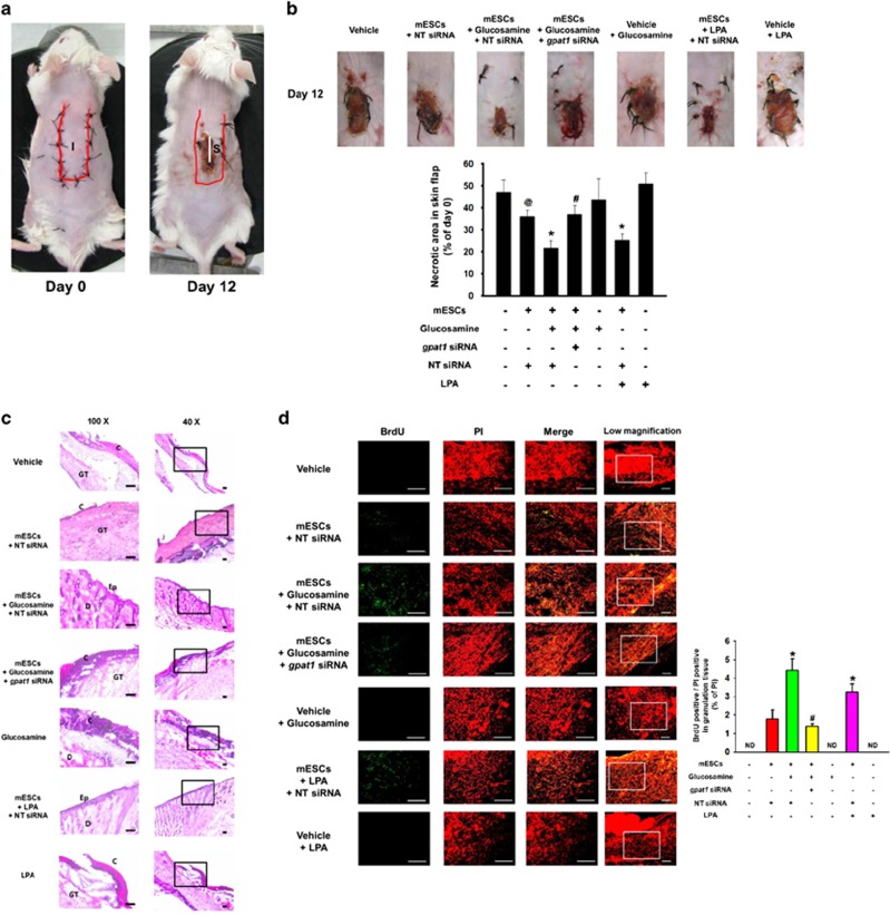
Role of GPAT1 in mESCs survival in the mouse skin flap model. Mouse skin flap surgery with BrdU-harbored mESCs transplantation was performed as described in Materials and methods section. (a) NT siRNA-transfected cells are injected in the center region (I) of the flap. At post-injection day 12, all tissue samples (S) including injection site in are excised and collected. (b) Representative gross images of skin flap were obtained at day 12 after flap surgery. Necrotic area in skin flap was analyzed by using ImageJ software. Error bars indicate a mean±S.E.M. n=5. @P<0.05 versus vehicle group, *P<0.05 versus mESCs group, and #P<0.05 versus mESCs with glucosamine group. (c) Tissue samples were stained with hematoxylin and eosin. Histological images shown in result are representative. Scale bars, 100 μm (magnification, × 40 and × 100). (d) BrdU was immunostained with BrdU specific antibody and PI for nuclear counting, and samples were visualized by using confocal microscopy. BrdU and PI-stained cells were analyzed by using MetaMorph software. Scale bars, 200 μm (magnification, × 100 and × 200). *P<0.05 versus mESCs group, and #P<0.05 versus mESCs with glucosamine group. C, crust; Ep, epidermis; D, dermis; GT, granulated tissue; ND, not detected

