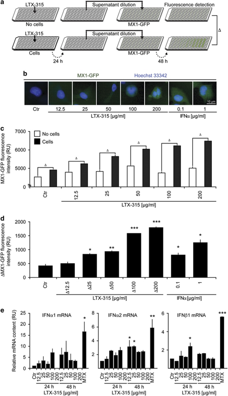
Figure 4.
Induction of a type-1 interferon response by LTX-315. (a–d) A schematic representation of the experimental design is shown in (a). LTX-315 was added at variable concentrations to culture media (without cells, above) or U2OS cell cultures (below). Recombinant IFN-α1 was used as a positive control. After 24 h, the culture supernatants were recovered and added to fresh cultures (1:16 dilution) of U2OS cells stably expressing GFP under the MX1 promoter (MX1-GFP). After an additional 48-h culture period, cells were fixed, counterstained with Hoechst 33342, and subjected to automated fluorescence microscopy and image analysis. Representative images are shown in (b), raw data of quantitations (means±S.D. of triplicates) in (b), and the subtraction of initially cell-free versus-cell-containing cultures in (d, e). Detection of type-1 interferons by RT-PCR. Cells were incubated as indicated with variable amounts of LTX-315 for distinct periods and then subjected to mRNA extraction and RT-PCR. Asterisks indicate significant differences (unpaired Student's t-test) with respect to untreated controls. *P<0.05; **P<0.01; ***P<0.001

