Figure 9.
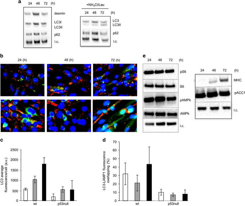
Autophagy is attenuated in the absence of p53 null cells and lysosomal activity is less efficient during myogenesis. (a) Left: WB analysis of desmin (53 kDa), LC3-I (18 kDa) and LC3-II (16 kDa) and p62 (62 kDa) performed on whole-cell extracts from p53 null cells during differentiation. Right: to measure the autophagic flux a mix of ammonium chloride and leupeptin (NH4Cl/Leu 20 mM/100 μM) was added to the medium 2 h before cell harvest. (b) Representative fluorescence microscopy images of p53 null muscle cells during differentiation. (Top) LC3 (green) and Lamp1 (red); (Bottom) p62 (red) and Beclin 1 (green). The nuclei were stained with DAPI. ImageJ quantification of fluorescence intensity of LC3 (c) and of LC3-Lamp1 (yellow area) versus LC3 (green area) (d) in wt and p53 null muscle cells was performed as described in Materials and methods section. Statistical analysis was performed by one-way ANOVA followed by Newman–Keuls test. *P=0.05, **P<0.01, ***P<0.001. (e) WB analysis of pS6, S6 (32 kDa), pAMPk and AMPK (62 kDa) (left) and MHC (200 kDa) and p-ACC (280 kDa) (right) in whole-cell extracts of p53 null muscle cells during differentiation (24, 48 and 72 h in DM); l.c.=loading control

