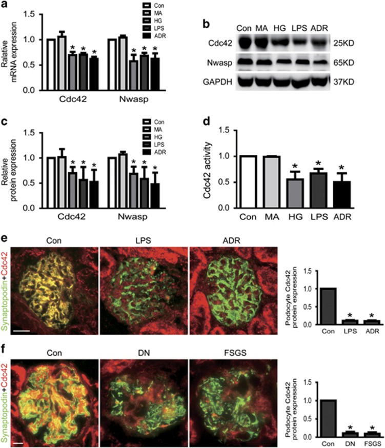
Figure 2.
Cdc42 and Nwasp were decreased in injured podocytes. (a) The mRNA expression of Cdc42 and Nwasp were decreased in HG, LPS or ADR-injured podocytes. (b, c) The protein expression of Cdc42 and Nwasp were decreased in HG, LPS or ADR-injured podocytes. (d) Cdc42 activity was decreased in HG, LPS or ADR-injured podocytes. (e) Representative micrographs of dual-color fluorescence staining of kidney glomeruli for Cdc42 (red) and synaptopodin (green) from control, LPS and ADR mice. Magnification × 400, scale bar=25 μm. (f) Representative micrographs of dual-color fluorescence staining of kidney glomeruli for Cdc42 (red) and synaptopodin (green) from control, DN and FSGS patients. Magnification × 400, scale bar=25 μm. All in vitro studies result in MA (as an osmotic control) and the Con group show hardly any difference (a–d). All data were from at least three independent experiments. *P<0.05 versus controls

