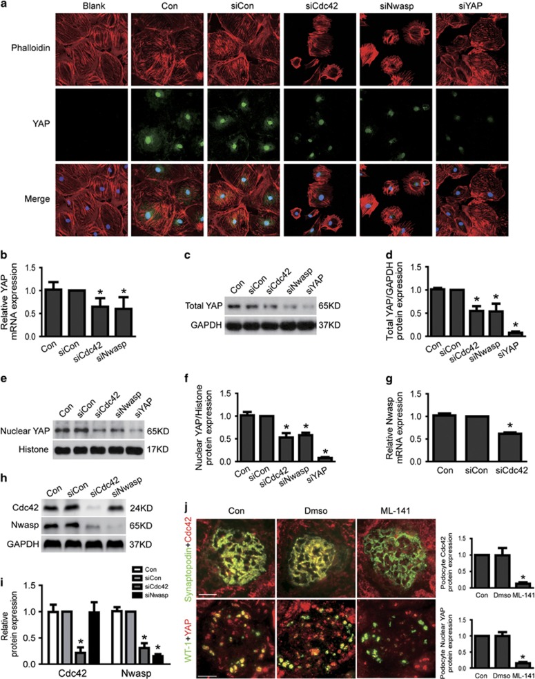
Figure 6.
Loss of Cdc42/Nwasp decreased stress fiber formation and YAP expression. (a) Typical confocal images of culture podocytes showing expression of YAP (green), Phalloidin-stained stress fiber (red) and DAPI-stained nuclei (blue). Compared to siCon, nuclear and cytoplasmic YAP and stress fiber were obviously reduced in Cdc42 siRNA or Nwasp siRNA-treated podocytes. (b) The mRNA expression of YAP was decreased in Cdc42 siRNA and Nwasp siRNA-treated podocytes. (c–f) The total and nuclear protein expression of YAP was decreased in Cdc42 siRNA or Nwasp siRNA-treated podocytes. (g) The mRNA expression of Nwasp was decreased in Cdc42 siRNA-treated podocytes. (h–i) Cdc42 and Nwasp protein expression were reduced to about 21.8%, 15.4% in Cdc42 siRNA and Nwasp siRNA-treated podocytes, respectively. Nwasp protein expression was obviously decreased in Cdc42 knockdown podocytes comparing to siCon, as Cdc42 protein expression was not changed in Nwasp knockdown podocytes. (j) On top: typical images of dual-color fluorescence staining of kidney glomeruli for synaptopodin (green) and Cdc42 (red) from control, Dmso and ML-141 (Cdc42-specific inhibitor) mice. Cdc42 was hardly seen in podocytes from ML-141 mice. Below: representative micrographs of dual-color fluorescence staining of kidney glomeruli for WT-1 (green) and YAP (red) from control, Dmso and ML-141 mice. Nuclear YAP was decreased in podocytes from ML-141 mice comparing to Con. Magnification × 400, scale bar=25 μm. All above data were from at least three independent experiments. *P<0.05 versus controls

