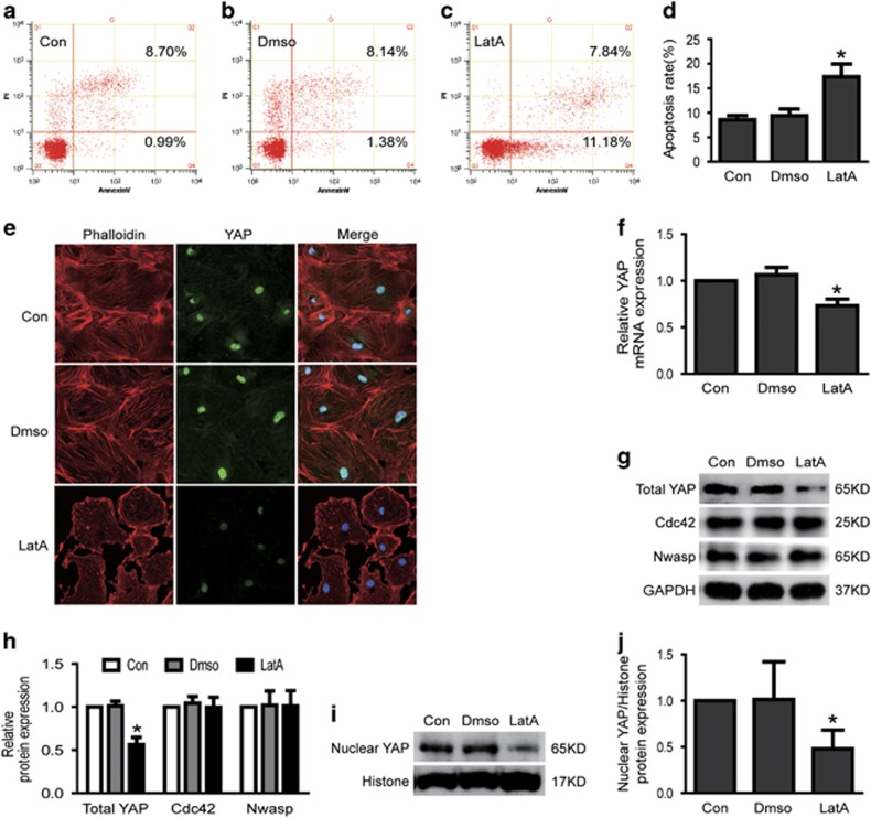
Figure 7.
Inhibited formation of stress fiber induced podocyte apoptosis and decreased YAP expression. (a–d) Podocytes were stained with Annexin V/PI for flow cytometry analysis. Cell apoptosis rate was significantly increased in LatA-(F-actin inhibitor, inhibited stress fiber formation) treated podocytes. (e) Typical confocal images of culture podocytes showing expression of YAP (green), Phalloidin-stained stress fiber (red) and DAPI-stained nuclei (blue). Compared to Con, nuclear and cytoplasmic YAP and stress fiber were obviously reduced in LatA-treated podocytes. (f) The mRNA expression of YAP was decreased in LatA-treated podocytes. (g–j) Total and nuclear protein expression of YAP was decreased in LatA-treated podocytes, but the protein expression of Cdc42 and Nwasp were not changed. All above data were from at least three independent experiments. *P<0.05 versus controls

