Figure 6.
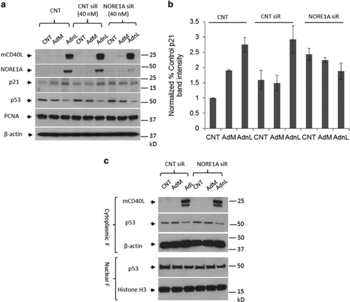
mCD40L-induced p21 expression is mediated by NORE1A protein. (a and b) EJ cells were transfected with 40 nM of either off-target siRNA or NORE1A siRNA or left untransfected as a negative control for 48 h. Cells were lightly trypsinzed and collected followed by infection with 100 MOI of either RAdMock (AdM) or RAdnCD40L (AdnL) or left uninfected as a negative control for further 24 h. Cells were lysed in situ and total protein lysates were examined for NORE1A, mCD40L, p21, p53, proliferating cell nuclear antigen and β-actin expression as a loading control (a). Expression levels of p21 were analysed by densitometry utilising the web-available ImageJ software and normalised to β-actin expression (b) results are average of two independent experiments ±S.D. (c) EJ cells were transfected with 40 nM of either off-target siRNA or NORE1A siRNA for 48 h. Cells were lightly trypsinzed and collected followed by infection with 100 MOI of either AdM or AdnL for further 24 h. Cytoplasmic and nuclear fractions were prepared and mCD40L, p53 and β-actin were examined in the cytoplasmic fraction, p53 and histone H3 as a loading control were examined in the nuclear fraction

