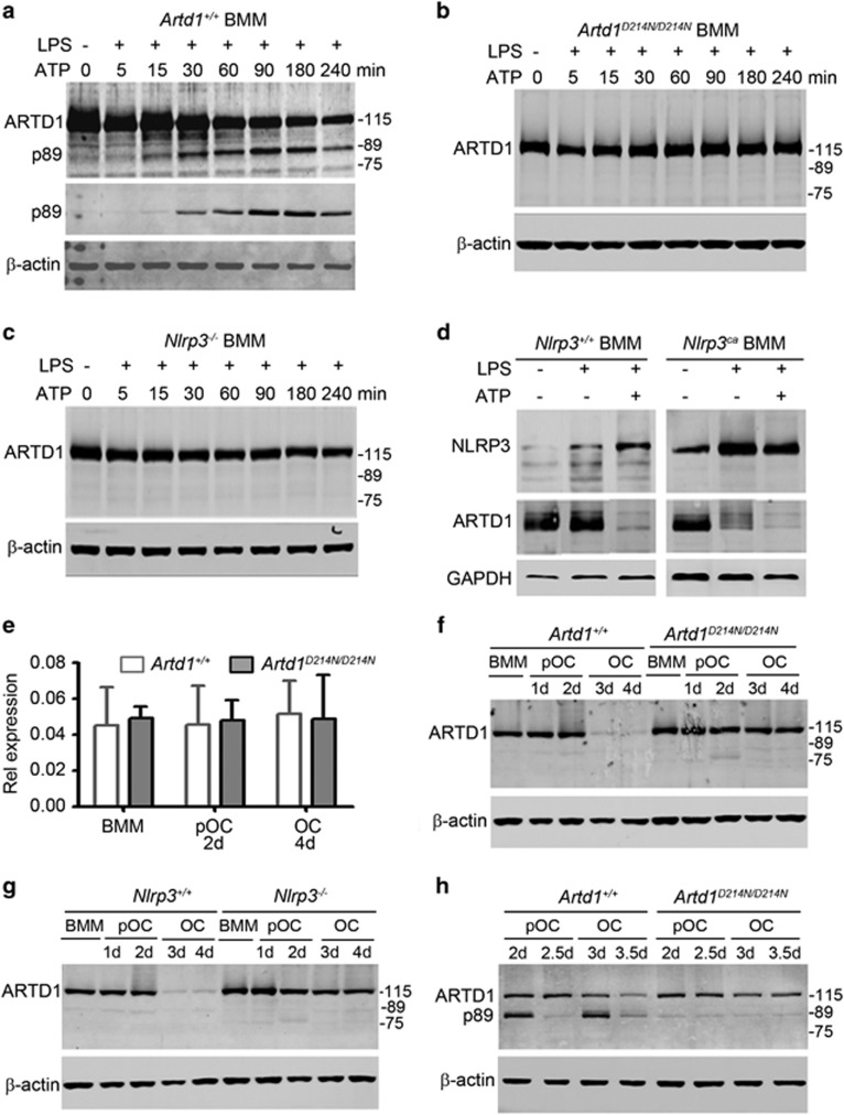
Figure 2.
Cleavage of ARTD1 at aspartate 214 is required for its degradation during osteoclastogenesis. BMM isolated from Artd1+/+ mice (a), Artd1D214N/D214N mice (b), Nlrp3−/− mice (c), WT mice or mice expressing constitutively activated NLRP3 inflammasome (Nlrp3ca, d) were treated with vehicle or 100 ng/ml LPS for 3 h, and exposed to vehicle or 5 mM ATP for the indicated times (min, minutes). Western blot analysis was carried out using ARTD1 antibody (top panel) or p89 ARTD1 antibody (middle panel, a). The lanes from the same membranes were cut and pasted in d. (e) WT and Artd1D214N/D214N BMM were incubated with 2% CMG (BMM) or 2% CMG and 100 ng/ml RANKL for 2 days (pOC, 2d) or 4 days (OC, 4d), and mRNA expression was analyzed by qPCR. (f) Analysis of ARTD1 degradation during OC formation from Artd1+/+ or Artd1D214N/D214N BMM. (g) Analysis of ARTD1 degradation during OC formation from Nlrp3+/+ or Nlrp3−/− BMM. (f) and (g) BMM were fed with 2% CMG or 2% CMG and 100 ng/ml RANKL every 2 days. (h) Analysis of ARTD1 cleavage and degradation during OC formation from BMM expressing ARTD1+/+ or ARTD1D214N. BMM were fed with 2% CMG and 100 ng/ml RANKL every day starting at day 1.5, and samples were analyzed 12 h later (day 2 or 3) or 24 h later (day 2.5 or 3.5). Samples were analyzed by Western blot; a nonspecific faint band around 89 kDa can be seen across samples (f–h). Data are representative of at least two independent experiments

