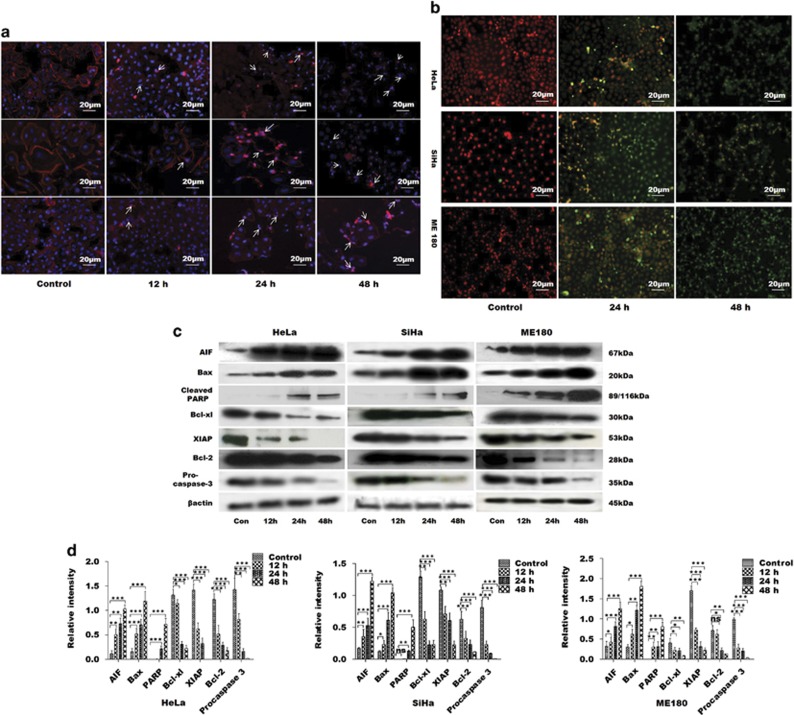
Figure 2.
GW627368X induces apoptosis in cervical cancer cells HeLa, SiHa and ME 180 in time-dependent manner. Cells treated with 9, 10 and 10 μM GW627368X for Hela, SiHa and ME 180, respectively, for indicated time periods, (a) stained with Rhodamine phalladoin and counter stained with DAPI and images captured at × 20 magnification. Nuclear fragmentation and disruption of actin cytoskeleton, characteristic of apoptotic cell death, was observed (b) TUNEL assay was performed and fluorescent images were captured. Time-dependent increase in TUNEL-positive green nuclei indicated induction of apoptosis, (c and d) Western blot analysis of apoptotic proteins, (c) Blots depicting expression profile of apoptotic and anti-apoptotic proteins in cervical cancer cells treated with GW627368X for indicated time periods compared with control treated with 0.1% DMSO, β-actin used as loading control (d) Quantification of protein expression by densitometry, normalized to β-actin, each representative of three different experiments each performed in triplicates, values presented as mean±S.D.; *P<0.05, **P<0.01, ***P<0.001

