Figure 4.
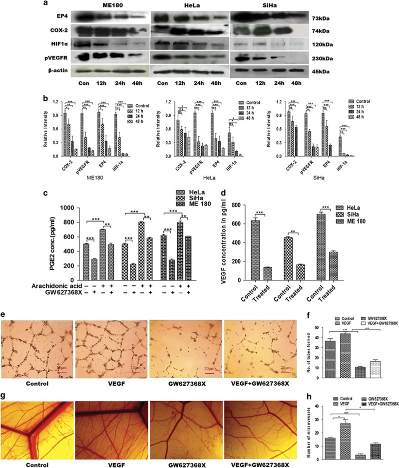
Anti-angiogenic potential of GW627368X. Cyclo-oxygenase/PGE2 axis is known to have prominent role in angiogenesis. (a) Western blot analysis of major players of COX-PGE2 axis in cervical cancer cell lines treated with GW627368X (9, 10 and 10 μM for Hela, SiHa and ME 180, respectively) for different time points and control treated with 0.1% DMSO. (b) Quantification of protein expression by densitometry, normalized against β-actin, each representative of three different experiments performed in triplicates, values presented as mean±S.D.; *P<0.05, **P<0.01, ***P<0.001. (c) Quantitative analysis of the PGE2 concentration in conditioned media of GW627368X treated cells (9, 10 and 10 μM for Hela, SiHa and ME 180, respectively), stimulated or unstimulated by 10 μM arachidonic acid by ELISA. Graphs representative of three different experiments each performed in triplicates, values presented as mean±S.D.; P<0.05, **P<0.01, ***P<0.001. (d) Quantitative estimation of the secretory VEGF concentration in conditioned media of GW627368X treated cells (9, 10 and 10 μM for Hela, SiHa and ME 180 respectively) by ELISA. Graphs representative of three different experiments each performed in triplicates, values mean±S.D.; P<0.05, **P<0.01, ***P<0.001. (e and f) In vitro tube formation assay. (e) HUVECs were seeded (7.5 × 10³ cells/well) into a 96-well tissue culture plate coated with 50 μl matrigel; VEGF (25 ng/ml), GW627368X (IC50 for 48 h/3)or both were added and incubated in HUVEC growth medium in 37 °C, 5% CO2 incubator. Tube formation was observed for 24 h and images were taken at × 10 magnification (f) Number of capillary-like structure formed by HUVECs in each condition was counted under light microscope after 24 h in four independent experiments. Data presented as mean±S.D., P<0.05, **P<0.01, ***P<0.001. (g and h) Chorioallantoic membrane (CAM) assays. (g) Photomicrographs representing developing chick embryos implanted with sterile filter disks loaded with serum-free media as control and serum-free media supplemented with VEGF (25 ng/ml), GW627368X (IC50 for 48 h/3) or both to observe the angiogenic response (h) The number of blood vessels formed in each test group was counted under stereo microscope and plotted to evaluate the extent of angiogenic response. Data presented as mean±S.D., P<0.05, **P<0.01, ***P<0.001 of three independent experiments performed in triplicates

