Figure 8.
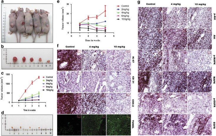
GW627368X hinders tumor progression in vivo. Subcutaneous tumor-bearing nude mice were treated with GW627368X orally ranging from 0 to 10 mg/Kg body weight, thrice a week, for 4 weeks. (a) Representative picture showing subcutaneous tumor (ME 180)-bearing nude mice of different treatment groups. (b) Representative picture showing excised tumors from corresponding treatment groups of ME 180 xenografts. (c) Graph representing change in tumor volume (group average; ME 180 xenografts) in each test group measured every week. Data presented as mean±S.D. (n=5), P=0.0125. (d) Representative picture showing tumors excised from different treatment groups of SiHa xenografts. (e) Graph representing change in tumor volume (group average; SiHa xenografts) in each test group measured every week. Data presented as mean±S.D. (n=5), P=0.0002. Paraffin-embedded excised tumors from different treatment groups of ME 180 xenografts were sectioned, processed and (f) In situ TUNEL assay to assess induction of apoptosis within tumor and IHC specific for COX-2, Ki67(proliferation marker) and CD31(anigiogenesis marker) were performed (g) Photomicrographs representing IHC performed for specific signaling proteins, pMAPK, MAPK, pAkt, Akt, pEGFR and EGFR

