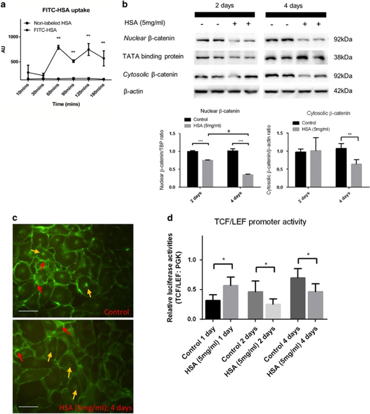
Figure 1.
β-Catenin expression is abrogated in protein overloaded HK-2 cells. (a) Albumin uptake measurement (n=3) in lysed HK-2 cells treated with FITC-human albumin/non-labeled HSA. (b) HK-2 cells were treated with 5 mg/ml HSA for 2 (n=6) and 4 (n=6) days. Immunoblotting assays of active β-catenin expression in the nuclear fraction and cytosolic fraction of the cell lysate. (c) Immunofluorescent staining of β-catenin in HSA-treated HK-2 cells for 4 days. Red arrows show the membrane-bound β-catenin and yellow arrows indicate nuclear β-catenin. (d) Luciferase assay of TCF/LEF reporter activities in HSA-treated or control HK-2 cells. Bar scale=25 μm. Graphs were expressed in mean± S.D. *P<0.05, **P<0.001, ***P<0.0001; #P<0.01

