Figure 3.
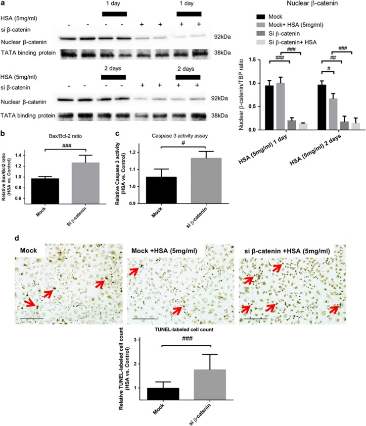
Loss of β-catenin/HSA stimulation promotes apoptosis in HK-2 cells. HK-2 cells were transfected with siRNA against β-catenin (si β-catenin) or mock siRNA (40 nM) for 2 and 3 days with or without addition of HSA (5 mg/ml) for 1 and 2 days. (a) Protein levels of nuclear β-catenin in transfected HK-2 cells treated with HSA for 1 day (n=3) and 2 days (n=6). (b) Bax/Bcl-2 gene expression in mock/si β-catenin transfected HK-2 cells under HSA stimulation for 2 days (n=6). (c) Caspase-3 activity in the corresponding groups of HK-2 cells after 2-day HSA treatment (n=3). (d) TUNEL-positive cells after 1-day HSA stimulation in HK-2 cells with or without si β-catenin transfection (n=6). Red arrows indicate apoptotic nuclei. Bar scale=25 μm. Graphs were expressed in mean± S.D. #P<0.05, ##P<0.001, ###P<0.0001

