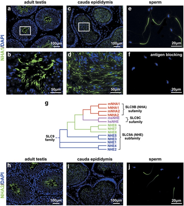
Figure 1.
NHA1 and NHA2 were specifically expressed in the principal piece of sperm tail. (a-f) Immunofluorescence staining of NHA1 in mouse testis (a and b), cauda epididymis (c and d) and sperm from cauda epididymis (e). Note the intensive green signal at principal piece of sperm tail. NHA1 antibody staining in the presence of competing immunogen (f). The nuclei are counterstained with DAPI (blue). (g) Phylogenetic tree displays the relationship between the NHA1 and the other NHEs. The tree was generated with GeneBee aligning the predicted open reading frames of each NHE. (h-j) Immunofluorescence staining of NHA2 in mouse testis (h), cauda epididymis (i) and sperm from cauda epididymis (j). Scale bar in a, c, h and i, 100 μm. Scale bar in b and d, 50 μm. Scale bar in e, f and j, 20 μm

