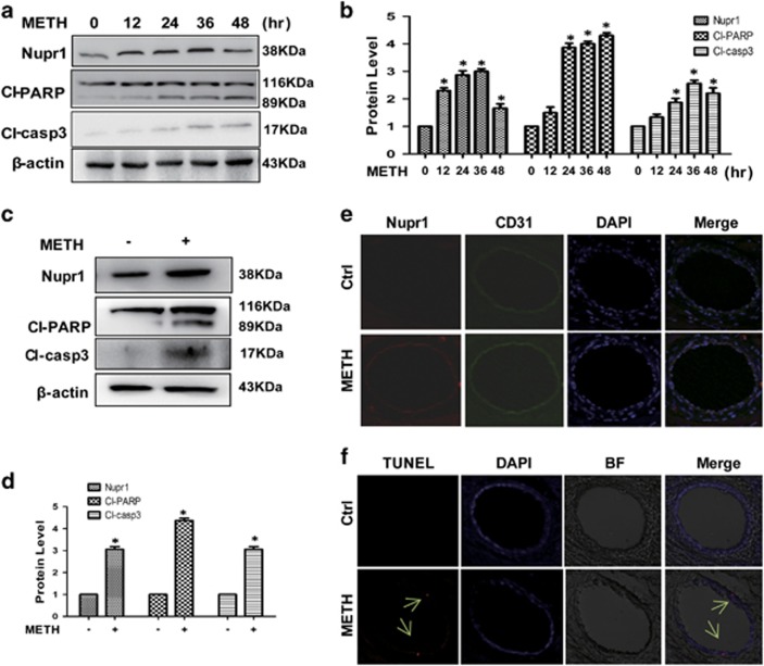
Figure 1.
Nupr1 expression is upregulated in endothelial cells after METH exposure in vitro and in vivo. (a) HUVECs were exposed to 1.25 mM METH for indicated times (0, 12, 24, 36 and 48 h). (c) CMECs were exposed to METH (0.5 mM) for 24 h. The protein expression of Nupr1, cleaved-PARP and cleaved-caspase-3 was determined with western blot (a and c) and quantitative analyses (b and d). Fold induction relative to cells treated with vehicle is shown. β-Actin was used as a loading control. *P<0.05 versus vehicle-treated cells. Data were analyzed with one-way ANOVA followed by LSD post hoc comparisons. Data represent mean±S.D. (n=3 replicates). (e and f) Male SD rats (n=5/group) were injected i.p. with saline or METH (15 mg/kg × 8 injections, at 12 h interval). The heart tissues were harvested at 24 h after the last dosing. Immunolabeling and confocal imaging analysis (e) showed elevated Nupr1 expression in the heart microvascular endothelial cells of METH-exposed rats compared with controls (Ctrl). TUNEL staining and confocal imaging analysis (f) was used to evaluate the endothelial cell apoptosis. Apoptotic cells were stained with TUNEL (Red). Nuclei were counterstained with DAPI (blue) and BF represents bright field

