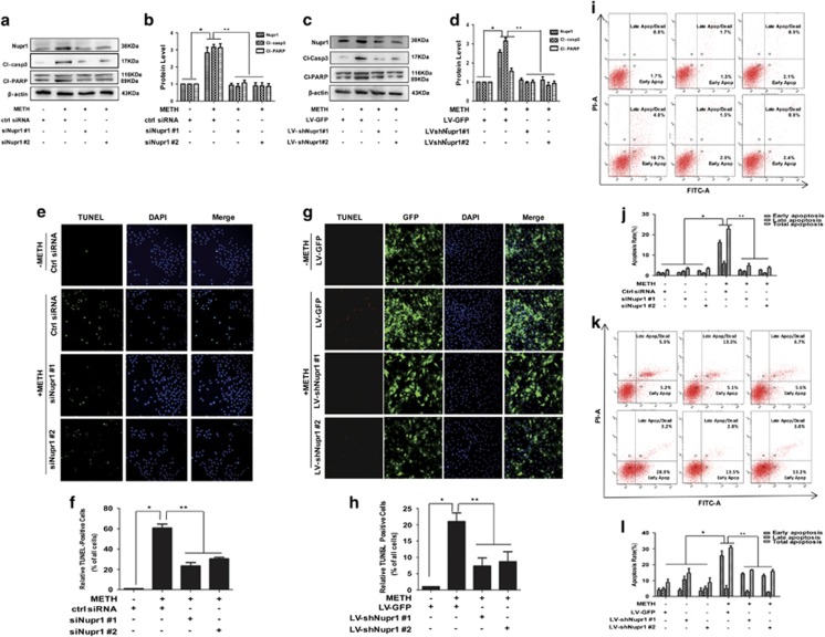
Figure 2.
Nupr1 is necessary for METH-induced apoptosis in HUVECs and CMECs. (a) Synthetic Nupr1 siRNAs effectively suppressed endogenous Nupr1 expression on HUVECs. HUVECs were transfected with siRNAs targeting Nupr1 or control siRNA for 48 h followed by METH (1.25 mM) treatment for 24 h. (c) Lentivrus-mediated Nupr1 shRNAs effectively suppressed endogenous Nupr1 expression in CMECs. CMECs were incubated with viral supernatants for 24 h followed by METH (0.5 mM) treatment for another 24 h. Western blot (a and c) and quantitative analyses were performed to evaluate the efficiency of Nupr1 knockdown (b and d), and the expression of apoptosis-related proteins (cleaved-PARP (Cl-PARP) and cleaved-caspase-3 (Cl-casp3)) after knocking down Nupr1 expression in HUVECs (b) and CMECs (d). Effects of suppressing Nupr1 expression on the apoptosis caused by 1.25 mM METH in HUVECs and by 0.5 mM METH in CMECs were assessed with TUNEL (e: green for HUVECs; g: red for CMECs) and flow cytometry ((i) HUVECs and (k) CMECs). (f and h) Quantitative analysis of the percentage of apoptotic cells using a standard cell counting method with the TUNEL assay. Apoptotic cells were stained with TUNEL (green for HUVECs (f) and red for CMECs (h)). Nuclei were counterstained with DAPI (blue). (j and l) Quantitative analysis of the effects of knocking down Nupr1 on apoptosis induced by METH using flow cytometry. Representative calculations from three independent replicates are shown. The percentage of apoptotic cells is presented as mean± S.D. (n=3, *P<0.01 versus the saline vehicle-treated control group; **P<0.01 versus the scrambled+METH group; one-way ANOVA)

