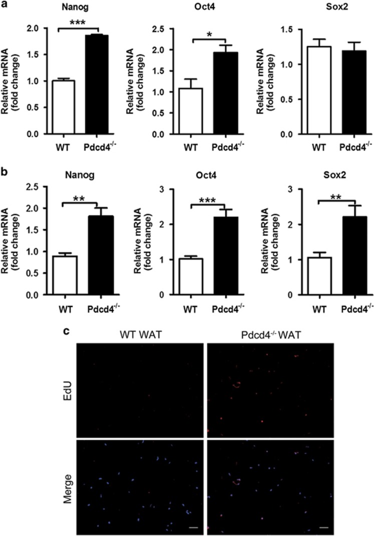
Figure 2.
Pdcd4 deficiency increases the stemness of ADSCs in WAT. (a) ADSCs from WT and Pdcd4−/− mice were isolated and expanded as described in Figure 1, the gene expression of stemness markers on ADSCs were measured by qPCR. Bars represent mean±S.E.M. *P<0.05, ***P<0.001. (b) The primary stromal vascular fraction was isolated from epididymal fat pads of WT and Pdcd4−/− mice (n=4 per group), the gene expression of stemness markers were examined by qPCR. Bars represent mean±S.E.M. **P<0.01, ***P<0.001. (c) WT or Pdcd4−/− mice (n=4) was intraperitoneally injected with 100 μg EdU. After 24 h, the epididymal fat pads were collected and paraffin-embedded sections were prepared. The incorporation of EdU in dividing cells was examined by fluorescence signals and visualized with fluorescence microscope. The original magnification is 100. Scale bar=100 μm

