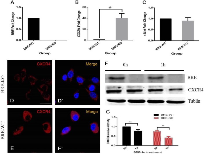
Fig. 10.
RT-qPCR analysis revealed that BRE-KO satellite cells expressed a significantly higher level of CXCR4 mRNAs (40 folds higher) than BRE-WT satellite cells (B, *P<0.05). (C) There was no significant difference in HGF receptors (c-Met) expression. (D–E′) Surprisingly, immunofluorescent staining indicated that BRE-WT satellite cells (E) were more intensely stained for CXCR4 expression than BRE-KO satellite cells (D). (F) Western blot analyses of CXCR4 were performed before and after 1 h SDF-1α treatment. (G) Statistical analysis revealed that CXCR4 was significantly degraded in both BRE-KO and BRE-WT satellite cells after 1 h SDF-1α treatment (**P<0.01). However, CXCR4 degradation in BRE-KO cells was significantly more rapid than in BRE-WT cell (50% vs 20%, respectively). Data are presented as mean±s.e.m. Scale bars=20 μm.

