Fig. 5.
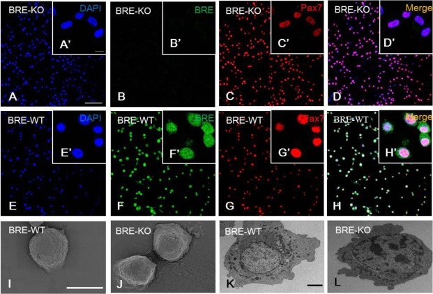
BRE-KO (A-D′) and BRE-WT (E-H′) satellite cells immunofluorescently stained with BRE and pax7 antibodies. The staining show BRE is expressed in the nucleus and cytoplasm of BRE-WT satellite cells (F). In the absence of BRE, the satellite cells are still capable of expressing pax7 (C). Under the scanning electron microscope, the morphology of both BRE-WT (I) and BRE-KO (J) satellite cells appeared rounded. Under the transmission electron microscope, the satellite cells show a high nuclear to cytoplasmic ratio in both BRE-WT (K) and BRE-KO (L) satellite cells. There was no morphological difference between the two groups. Scale bars, 100 μm for A-H, 10 μm for A′-H′, 100 μm for I-J and 20 μm for K-L.

