Fig. 1.

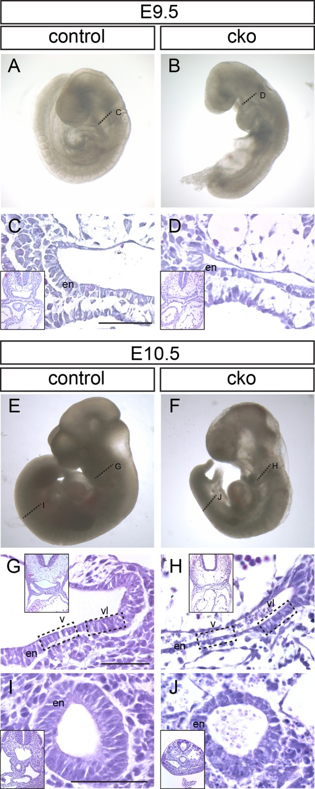
Consequences of Cdc42 ablation following tamoxifen administration at E8.5. (A-D) Control (A) and Cdc42-conditional knockout (cko; B) embryos from the same litter collected at E9.5. (C,D) Sections through the foregut (fg) region of embryos at the locations indicated by the dotted lines in panels A and B. (E-J) Control (E) and conditional knockout (F) embryos collected at E10.5. Dotted lines indicate the location of sections through the foregut (G,H) and hindgut (I,J) regions (hg). Boxed regions in G and H indicate the ventral (v) and ventral-lateral (vl) foregut endoderm (en) referred to in subsequent figures. Low magnification images corresponding to each panel are shown as insets. Scale bar=100 μm.
