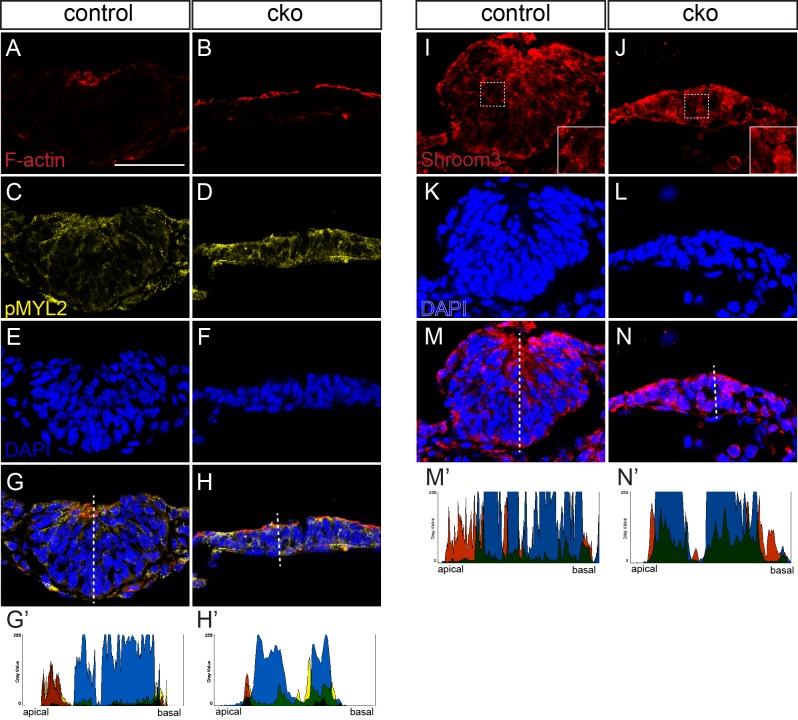
Fig. 7.
Impaired apical constriction in Cdc42-deficient thyroid buds. (A-H) Phalloidin staining of F-actin (A,B) and immunostaining of phosphorylated myosin II light chain (C,D) in control and conditional knockout (cko) thyroid buds. (I-N) Immunostaining for SHROOM3 in control and conditional knockout thyroid buds. Insets in I and J show higher magnification of the corresponding boxed region in the main panel. Specimens were counterstained with DAPI to reveal the nuclei (E,F,K,L). (G-H′,M-N′). Labelling was merged in G,H,M,N, and fluorescence intensity was measured on the representative specimen shown in G′,H′,M′,N′, in the plane of the dotted lines in panels G,H,M,N. Colors shown in the charts indicate the protein stained with the same color in the corresponding fluorescence image.

