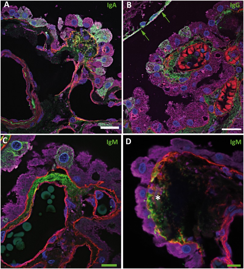
FIGURE 4.
Immunoglobulins in the choroid plexus. Confocal micrographs showing immunoglobulins in green, basement membrane (collagen IV) in red, epithelial cells (transthyretin) in purple, and cell nuclei (DAPI) in blue. (A) IgA (green) is seen in the stroma, focally colocalizing in the subepithelial basement membrane (yellow), and in the cytoplasm of epithelial cells. (B) IgG (green) is evident in the stroma and in some epithelial cells near IgG-positive ependymal cells (green arrows). (C) IgM (green) is focally evident in the stroma and in some epithelial cells. (D) IgM deposition in a concretion (asterisk), which is associated with a defect in the overlying subepithelial basement membrane. There is focal basement membrane thickening within which there is colocalization with IgM (yellow). Some basement membrane material is also seen within the concretion. Scale bars: green = 10 µm; white = 20 µm.

