Fig. 4.
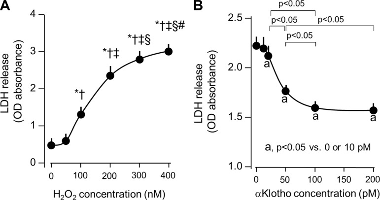
Cytoprotection by αKlotho in an in vitro hydrogen peroxide (H2O2) model of oxidative damage. A: dose-response of H2O2 on lactate dehydrogenase (LDH) release as a marker of cell death. A549 lung epithelial cells were incubated with the indicated H2O2 dose for 4 h. *P < 0.0001 vs. 0 nmol/l H2O2, †P < 0.0001 vs. 50 nmol/l H2O2, ‡P < 0.001 vs. 100 nmol/l H2O2, §P < 0.05 vs. 200 nmol/l H2O2, #P < 0.05 vs. 300 nmol/l H2O2. B: dose-response of purified recombinant αKlotho on H2O2-induced A549 cell death. aP < 0.05 vs. 0 or 10 pM αKlotho concentration. Average of 3 independent experiments (means ± SD). Statistical significance was assessed by ANOVA.

