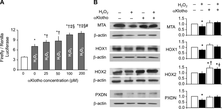
Fig. 6.
Activation of nuclear-factor erythroid derived 2 transcription factor (Nrf2) pathway in lung epithelial cells by αKlotho. A: A549 cells were transfected with an antioxidant responsive element (ARE)-luciferase reporter and were treated with the stated concentration of recombinant αKlotho. Activation of ARE is expressed as firefly luciferase luminescence ratio to the control Renilla. Average of 3 independent experiments (means ± SD). Statistical significance was assessed by ANOVA. *P < 0.0001 vs. untreated cells, †P < 0.001 vs. cells treated with 200 nM H2O2 and 0 pM αKlotho, ‡P < 0.0001 vs. cells treated with 200 nmol/l H2O2 and 25 pM αKlotho, §P < 0.0001 vs. cells treated with 200 nM H2O2 and 50 pM αKlotho, #P < 0.05 vs. cells treated with 200 nM H2O2 and 100 pM αKlotho. B, left: selected antioxidant protein expressions were quantified by immunoblot. MTA, methalothionine; HOX1 and HOX2, heme oxygenase 1 and 2; PXDN, peroxidasin. β-Actin served as loading control. B, right: densitometry of antioxidant levels was normalized to β-actin and expressed as a ratio to control (no H2O2, no αKlotho). Average of 3 independent experiments (means ± SD). Statistical significance was assessed by ANOVA. *P < 0.05 vs. control, †P < 0.05 vs. 200 mM H2O2 and no αKlotho, ‡P = 0.065 compared with 200 mM H2O2 and no αKlotho.

