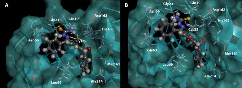
Figure 6.
Molecular docking of analogues 1 and 32 within the active site of cathepsin L. (A) Analogue 1 and (B) analogue 32 are shown in ball and stick mode (C, gray; H, white; O, red; N, blue; S, yellow; F, light blue; Br, brown). Cathepsin L is shown in ribbon mode with a transparent molecular surface (Green) and enzyme active site amino acid residues are labeled and shown in stick mode (C, gray; H, white; O, red; N, blue; S, yellow).

