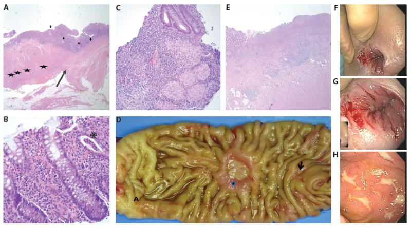
Figure 1.

Pathology and Colonoscopy Images
Panels A-C are pathology slides from case #5. Panel A Redo pull through resection. Long arrow marks the anastomotic line. Ganglion cells are noted between the muscular layers in the proximal segment (left, stars). The distal mucosal sleeve is aganglionic (right). Non necrotizing epithelioid granulomas are noted on both sides of anastomosis (short arrow). Panel B Colonic biopsy shows diffuse chronic inflammation of the lamina propria and focal crypt abscess (asterisk). Panel C Numerous epithelioid granulomas seen in the colonic biopsies.
Panels D-E are images from case #6. Panel D Small bowel resection shows two chronic ulcers, small (arrow) and large (star) proximal to the anastomotic site (A). Panel E Deep ulceration and underlying transmural chronic inflammation and fibrosis resembling Chron disease.
Panels F-H are initial colonoscopy images of case #4. Panel F is a gross image of perianal disease prior to anti-TNF-α treatment. Panel G is a closer image of the same perianal lesions. Panel H is an image of his aphthous ulcers.
