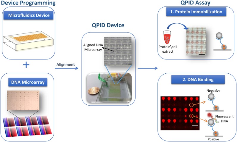
Figure 1.
QPID system overview. QPID experiments are programmed by aligning and bonding an integrated microfluidic device containing thousands of micromechanical valves (upper left) with a DNA microarray (lower left). Each color in the microarray has a gradient and represents a different DNA sequence at different concentrations. A typical QPID Device (middle) containing 32 different oligonucleotides at 32 different concentrations in four independent identical blocks is ready for performing 4096 individual quantitative binding experiments. Zoom in on a device layout shows the DNA microarray locked within the microfluidic chambers (upper middle). 1. We express the TFs in tubes using in vitro transcription and translation. We load the proteins or cell extracts (with over expressed proteins) onto the QPID device and immobilize them to the surface (upper right). 2. In the DNA binding assay (bottom right), the fluorescent DNA oligonucleotides are incubated with the TFs, MITOMI is performed and fluorescent images are taken. We measure the affinity of the TFs to each of the oligonucleotides at equilibrium and calculate the dissociation constant.

