Figure 3.
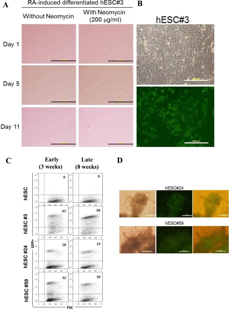
Functionality of transgenes in LINE-1-targeted hESCs. (A) Functional test for UTF1 promoter-driven neomycin resistance. hESC clone #3 was induced to differentiate by retinoic acid (RA) treatment and cultured in differentiation medium with or without neomycin for up to 12 days. In differentiated cells, cell death was evident from day 5, following neomycin treatment (bottom panels) while cells without neomycin treatment continued to grow (top panels). Magnification of 4×, Scale bars, 2 mm. (B) GFP expression in targeted hESC clones. A representative phase-contrast light micrograph (top panel) and fluorescence micrograph showing stable GFP expression (bottom panel) in hESC#3. Magnification of 10x, Scale bars, 500 μm. (C) FACS analysis of targeted clones. Dot plots representing GFP+ cells (upper right quadrant) and GFP− cells (lower right quadrant) for untargeted hESCs (parental cells) and targeted hESC clones #3, #24 and #59 after 3 weeks (early) and 8 weeks (late) of culturing the cells. (D) Cardiac differentiation of targeted hESC clones. Representative photomicrographs of cardiomyocytes generated from hESC clones #24 and #59. Light micrographs (left) and fluorescence micrographs showing EGFP expression in differentiated cells (middle). Overlay of light and fluorescence micrographs (right). Pulsating EGFP-positive cardiomyocytes could be seen at day 7 post induction. Movies of beating cardiomyocytes (Supplementary Movies 1–3).

