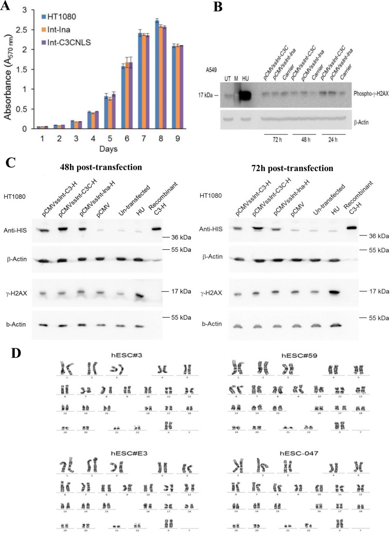
Figure 5.
Int-C3CNLS does not induce DNA damage or cytotoxicity. (A) MTT-based cell proliferation assays were performed to assess effects on cell proliferation rates upon Int expression in human cell lines. HT1080 cells untransfected (HT1080), and FACS sorted GFP+ cells obtained after co-transfection of pCMV-EGFP with either pCMVssInt-Ina (INA; expressing inactive integrase) or pCMVssInt-C3CNLS (C3CNLS) were analyzed for the effect on cellular proliferation using the colorimetric MTT assays over the indicated time course. Data show the mean of triplicates and standard deviation of a representative experiment. n = 2. (B) Western blot analysis to determine phospho-γ-H2AX levels to assess DNA damage induced by expression of Int in A549 cells. Cell lysates prepared at time points of 24, 48 and 72 h (post transfection) from A549 cells transfected with pCMVssInt-Ina or pCMVssInt-C3C (expressing Int-C3CNLS) and from control cells treated with the carrier (Lipofectamine2000 Transfection reagent) were subjected to western blot analysis using antibodies against phospho-γ-H2AX (top panel). UT, untreated cells as negative control; HU, cells treated with hydroxy urea (10 mM for 24 h) as positive control; M, Marker lane. β-actin was used as loading control (bottom panel). (C) Western blot analysis to determine phospho-γ-H2AX levels to assess DNA damage induced by expression of Int in HT1080 cells. Forty-eight hours post transfection, top and 72 h post transfection, bottom. Lysates from HT1080 cells transfected with pCMV vector, plasmids expressing 6xHIS-tagged Inactive integrase (pCMVssInt-Ina-H), 6xHIS-tagged Int-C3 (pCMVssInt-C3-H), 6xHIS-tagged Int-C3-CNLS (pCMVssInt-C3C-H) were analyzed by western blotting with anti-HIS tag antibodies and phospho-γ-H2AX antibodies. UT, untreated cells; HU, cells treated with hydroxy urea (10 mM for 24 h) as positive control; C3-H, purified recombinant HIS-tagged Int-C3. HIS-tagged Int variants were detected at the expected size of 40 kDa in lysates from cells transfected with the integrase expression plasmids. There was no detectable induction of phospho-γ-H2AX upon expression of Int-C3-H or Int-C3CNLS-H compared to inactive Int-expressing cells and HU-treated cells. β-Actin protein levels were determined as loading controls. (D) Karyotyping to verify chromosomal stability. The targeted hESC lines hESC#3, hESC#59, hESC#E3 and the parental hESC-047 were karyotyped by G-banding of metaphase chromosomes. A representative karyotype (from 20 scored and five analyzed GTG-banded cells) for each cell line is shown. Results indicated no apparent chromosomal abnormalities in the tested cell lines.

