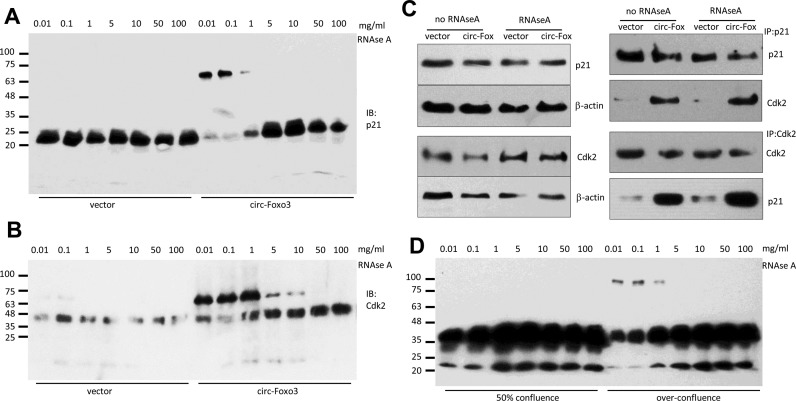
Figure 7.
CDK2 and p21 protected the circ-Foxo3 binding site. (A and B) The lysates prepared from circ-Foxo3- and mock-transfected cells were incubated with different concentrations of RNAse A followed by native gradient gel electrophoresis. The gels were subject to Western blotting probed with antibodies against p21 (A) or CDK2 (B). (C) Cell lysates from NIH3T3 cells transfected with circ-Foxo3 and mock control were treated with or without RNAse A (0.1 mg/ml), and subject to western blotting. While treatment with RNAse A did not affect levels of CDK2 and p21 (left), anti-p21 antibody was able to pull-down Cdk2 and anti-CDK2 antibody was able to pull down p21 in the circ-Foxo3-transfected cells with or without RNAse A treatment. (D) The lysates prepared from NIH3T3 cells grown to 50% conference and over-confluence were incubated with different concentrations of RNAse A followed by native gradient gel electrophoresis. The gels were subject to western blot probed with antibody against p21 and then against Cdk2.

