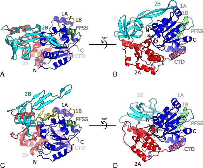
Figure 1.
Overall structure of BsPif1. Domains are colored as follows: 1A (blue), 1B (orange), 2A (red), 2B (cyan) CTD (pink), PFSS (green). (A) BsPif1 with AMPPNP (space group P212121). The nucleotide binding site is in the front with the nucleotide shown as ball-and-stick. (B) The view is rotated by 90°. (C) BsPif1 with ADP-AlF3-ssDNA (space group P3121). ssDNA is colored in yellow. BsPif1 is in the same orientation as in (A) with domains 1A superimposed. (D) Rotated view by 90° of BsPif1 with ADP-AlF3-ssDNA.

